Manal A Awad1 ![]() ,
Awatif A Hendi2,
Khalid MO Ortashi3,
Reem A Alotaibi4,
Maha Sh Sharafeldin5
,
Awatif A Hendi2,
Khalid MO Ortashi3,
Reem A Alotaibi4,
Maha Sh Sharafeldin5
For correspondence:- Manal Awad Email: mawad@ksu.edu.sa
Received: 28 November 2015 Accepted: 16 March 2016 Published: 30 April 2016
Citation: Awad MA, Hendi AA, Ortashi KM, Alotaibi RA, Sharafeldin MS. Characterization of silver nanoparticles prepared by wet chemical method and their antibacterial and cytotoxicity activities. Trop J Pharm Res 2016; 15(4):679-685 doi: 10.4314/tjpr.v15i4.2
© 2016 The authors.
This is an Open Access article that uses a funding model which does not charge readers or their institutions for access and distributed under the terms of the Creative Commons Attribution License (http://creativecommons.org/licenses/by/4.0) and the Budapest Open Access Initiative (http://www.budapestopenaccessinitiative.org/read), which permit unrestricted use, distribution, and reproduction in any medium, provided the original work is properly credited..
Purpose: To investigate the efficiency of silver nanoparticles synthesized by wet chemical method, and evaluate their antibacterial and anti-cancer activities.
Methods: Wet chemical method was used to synthesize silver nanoparticles (AgNPs) from silver nitrate, trisodium citrate dehydrate (C6H5O7Na3.2H2O) and sodium borohydride (NaBH4) as reducing agent. The AgNPs and the reaction process were characterized by UV–visible spectrometry, zetasizer, transmission electron microscopy (TEM) and scanning electron microscopy (SEM) equipped with energy dispersive spectroscopy (EDS). The antibacterial and cytotoxic effects of the synthesized nanoparticles were investigated by agar diffusion method and MTT assay respectively.
Results: The silver nanoparticles formed were spherical in shape with mean size of 10.3 nm. The results showed good antibacterial properties, killing both Gram-positive and Gram-negative bacteria, and its aqueous suspension displayed cytotoxic activity against colon adenocarcinoma (HCT-116) cell line.
Conclusion: The findings indicate that silver nanoparticles synthesized by wet chemical method demonstrate good cytotoxic activity in colon adenocarcinoma (HCT-116) cell lines and strong antibacterial activity against various strains of bacteria.
Introduction
Nanotechnology is probably one of the fastest developing sciences over the last few years. It is an interdisciplinary science that connects knowledge of biology, chemistry, physics, material science, engineering, pharmacology, and medicine. The products of nanotechnology, nanoparticles, have a size < 100 nm and, due to their small size, have very high bioactivity [1].
Metal nanoparticles can be prepared and stabilized by physical and chemical methods [2]. Numerous approaches had been used to prepare silver nanoparticles (AgNPs). Examples of physical technique to prepare AgNPs are photochemical synthesis, laser ablation, phase transfer processes, microemulsion, microwave treatment [3], which had been used for preparation in both solution and solid phases of silver nanoparticles. Chemical methods are widely used to produce nanostructured materials due to their straight forward nature and their potential to produce large quantities of the final product. The particle sizes of the nanoparticles can be controlled by systematically adjusting the reaction parameters, such as time, temperature [4], and the concentration of reagents and stabilizing agents [5]. A variety of chemical approaches have also been utilized to produce silver nanoparticles with different size distribution and different shapes [6].
Unique interactions with bacteria and virus have been demonstrated by silver nanoparticles of certain size ranges and shapes [7]. The antimicrobial properties of silver nano have been applied widely to biomedical devices, water purification, clothing, cosmetics and other numerous household products [8]. Cancer is an abnormal type of tissue growth in which the cells exhibit an uncontrolled division, relatively in an autonomous fashion, leading to a progressive increase in the number of dividing cell.
There is increasing demands for anticancer therapy. In vitro cytotoxicity testing procedures reduces the use of laboratory animals and hence use of cultured tissues and cells have increased. The discovery and identification of new antitumor drug with low side effects on immune system has become an essential goal in many studies of immuno-pharmacology. With this aim, many medically relevant nanoparticles such as silver nanoparticles (AgNPs) were investigated for their cytotoxicity aspect. AgNPs showed different degrees of in vitro cytotoxicity [9].
This study targets synthesis of silver nanoparticles using wet chemical method, the synthesized AgNPs as antibacterial and anti-cancer agent is also investigated.
Methods
Materials
Silver nitrate (AgNO3, 99.99 %), trisodium citrate dihydrate (C6H5O7Na3.2H2O, 99.99 %), sodium borohydride (NaBH4, 99.99 %), were purchased from Sigma - Aldrich, USA. Deionized water was used throughout the experiment. Gram positive and gram negative bacteria test was done in the Central lab, Women Section, King Saud University. Dimethyl sulfoxide (DMSO) and crystal violet were purchased from Sigma (St. Louis, Mo., USA). DMEM, HEPES buffer solution, L-glutamine and gentamycin were purchased from (Bio Whittaker ® Lonza, Belgium).
Preparation of silver nanoparticles
To synthesize silver nanoparticles (AgNPs), wet chemical method was used. The spherical AgNPs were prepared according to the procedure reported by Fang et al [10]. In this method, AgNPs were synthesized by using NaBH4 as reducing agent. An aqueous solution of trisodium citrate (0.5 mL, 6 mM) was added into a flask containing (50 mL of deionized water), and then, an aqueous solution of AgNO3 (1 mL, 1 mM) was added. Freshly prepared NaBH4 aqueous solution (0.5 mL, 10 mM) was quickly added, and the suspension immediately turned a light yellow color. After 10 s, the suspension changed to a darker yellow or brown color after reaction had proceeded for another 20 second.
Characterization of AgNPs
UV-visible spectroscopy analyss was carried out at a wavelength range 190 - 1100 nm in a Perkin Elmer UV-visible spectrometer Lambda 25, PerkinElmer, United Kingdom. The mean size of the silver nanoparticles was analyzed using Zetasizer, (Nano series, HT Laser, ZEN3600 from Molvern Instrument, UK), while transmission electron microscopy (TEM, JEM-1011, JEOL, Japan) was employed to determine the size, shape and morphologies of formed synthesized silver nanoparticles. Energy dispersive spectrometer (EDS) analysis was performed for the confirmation of elemental silver. Elemental analysis on single particles was carried out using Oxford Instrument, Incax-act, equipped with scanning electron microscopy (JEOL-FE SEM).
Antibacterial assay
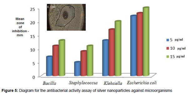
Antimicrobial activity of nanosilver was determined using disc diffusion assay method. Pure culture of Escherichia coli, Klebsiella pneumonia (gram negative), and Staphylococcus, Bacillis subtilis (gram positive) of bacteria were used. The sterile discs were dipped in nanosilver (5, 10, 15 μg/mL) and placed in the nutrient agar plate and kept for incubation at 37 oC for 24 h. Upon inhibitory activity, a zone of clearing around the wells was observed. The experiments were repeated 3 times and mean values of zone diameter were determined [11].
Evaluation of cytotoxic activity
Colon adenocarcinoma (HCT-116) cell lines were used. The tested human carcinoma cell line was obtained from the American Type Culture Collection (ATCC, Rockville, MD).
Chemicals
Staining solution
Crystal violet stain (1 %) is composed of 0.5 % (w/v) crystal violet and 50 % methanol then made up to volume with distilled water and filtered through a Whatmann No. 1 filter paper.
Cytotoxicity assay
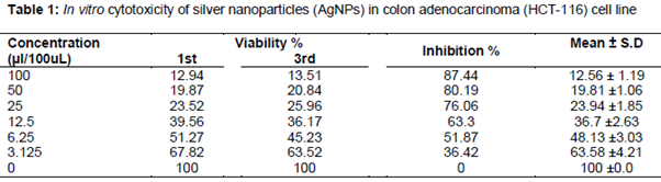
The cells were propagated in Dulbecco's modified Eagle's medium (DMEM) supplemented with 10 % heat-inactivated fetal bovine serum, 1 % L-glutamine, HEPES buffer and 50 μg/mL gentamycin. All cells were maintained at 37 oC in a humidified atmosphere with 5 % CO2 and were sub-cultured two times a week. Cell toxicity was monitored by determining the effect of the test samples on cell morphology and cell viability. For cytotoxicity assay, the cells were seeded in 96-well plate at a cell concentration of 1 x 10⁴ cells per well in 100 µL of growth medium. Fresh medium containing different concentrations of the test sample was added after 24 h of seeding. Serial two-fold dilutions of the tested chemical compound were added to confluent cell monolayers dispensed into 96-well, flat-bottomed microtiter plates (Falcon, NJ, USA) using a multichannel pipette. The microtiter plates were incubated at 37 oC in a humidified incubator with 5% CO2 for a period of 48 h. Three wells were used for each concentration of the test sample. The little percentage of DMSO present in the wells (maximal 0.1 %) was found not to affect the experiment. After incubation of the cells for 24 h at 37 oC, various concentrations of sample (100, 50, 25, 12.5, 6.25, 3.125 µl/100 uL) were added, and the incubation was continued for 48 h and viable cells yield was determined by colorimetric method. In brief, after the end of the incubation period, media were aspirated and crystal violet solution (1 %, 50 µl) was added to each well and allowed to stand for at least 30 min. The stain was removed and the plates were rinsed using tap water until all excess stain was removed. Glacial acetic acid (30 %, 100 uL) was then added to all the wells, mixed thoroughly, and then the absorbance of the plates was measured after gentle shaking in a microplate reader (TECAN, Inc.), using a test wavelength of 490 nm. All the results were corrected for background absorbance detected in wells without added stain. All experiments were carried out in triplicate. The cell cytotoxic effect of each tested compound was calculated [12,13].
Results
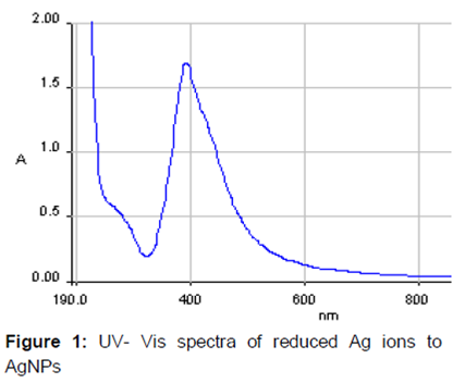
After the addition of fresh NaBH4 to aqueous solution of 1 mM AgNO3 and 6 mM trisodium citrate, the color changed immediately from colorless to yellowish brown in about 30 s. Formation of AgNPs was confirmed using UV–visible spectral analysis. The UV-vis spectrum exhibited an absorption band at 392.37 nm ().
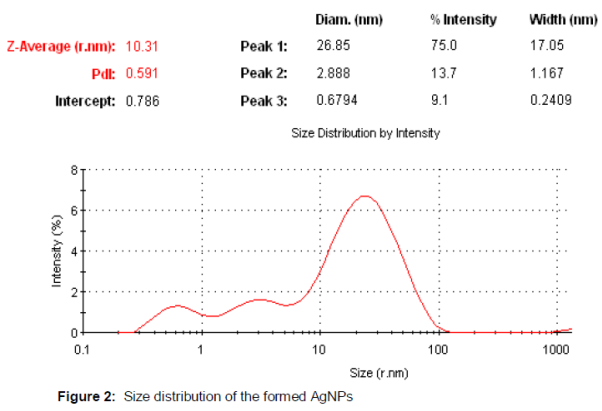
The results of dynamic light scattering (DLS) Zetasizer showed non-homogeneous AgNPs with mean particle size of 10.31 nm which can be observed in the appearance in three peaks ().
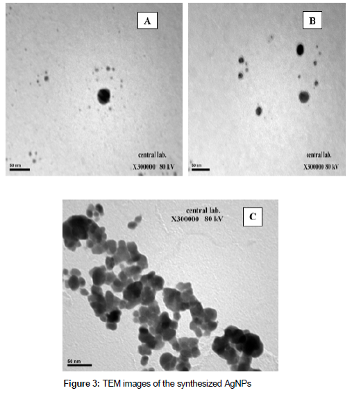
The results of transmission electron microscopy (SEM) show that the silver nanoparticles were irregular in shape with some spherical ().
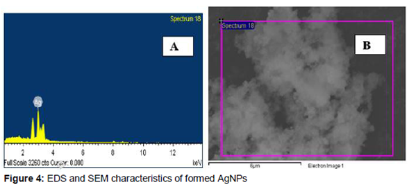
Energy dispersive x-ray analysis (EDS) revealed strong signal in the silver region and confirms the formation of silver nanoparticles (A). Morphological examination showed that the AgNPs agglomerated and has crystal and irregular morphology (B).
The results of AgNPs suspension tested against different gram positive (Staphylococcus aureus, Bacillis subtilis) bacteria and gram negative (Escherichia coli, Klebsiella pneumonia) bacteria show that AgNPs exhibited high toxicity against Escherichia coli, Klebsiella and low toxicity against Staphylococcus aureus. The data are shown in .
The results obtained from MTT assay after 48 h of incubation indicate that that AgNPs showed significant effect on HCT-116 with IC50 of 5.87 µl/100 uL ().
Discussion
The color change from colorless to yellowish brown when 1 mM solution of AgNO3 was added to the 6 mM trisodium citrate was due to excitation of surface plasmon vibrations [14]. Metal nanoparticles such as silver and gold have free electrons, which gives rises to surface plasmon resonance (SPR) absorption band [15]. The UV-vis spectrum of silver nanoparticles produced by wet chemical method exhibited an absorption band at 392.37 nm which is a typical plasmon band, suggesting the formation of silver nanoparticles. The results of dynamic light scattering (DLS) showed non homogeneous AgNPs with average particle size of 10.31 nm. The appearance of the three peaks in shows the polydispersity of nanoparticles which indicates that the nanoparticles may not be stable over a long storage period, and also that there are different sizes of the AgNPs in the suspension of the nanoparticles. TEM images recorded at high magnification morphology of the synthesized silver nanoparticles indicates that some of nanoparticles were spherical in shape with a smooth surface morphology while other were aggregated and irregular shaped. These results were suitable with the zetasizer results and UV–visible spectroscopic analysis. The energy dispersive X-ray analysis (EDS) revealed strong signal in the silver region and confirms the formation of silver nanoparticles, which illustrates that optical absorption peak was observed approximately at 3 keV, This is typical for the absorption of metallic silver nanostructure due to surface plasmon resonance (Morones et al, 2005). The morphology were carried out on the FE-SEM images showed the silver nanoparticles were agglomerated and has crystal line and irregular morphology, these result agreed with TEM results.
The effect of silver nanoparticle was more in Gram negative bacteria than Gram positive. This could be due to the fact that the relative abundance of negative charges on Gram negative bacteria assists the interaction between the nanoparticles and the cell wall [16]. Furthermore, the antibacterial properties shown above, allows us to confirm that our results are similar to those of Morones et al [17] who reported that small size nanoparticles may pass through cell membranes generating cell malfunction . Also, Choia et al has reported that the positive charge on the Ag ion is crucial for its antimicrobial activity through the electrostatic attraction between negative charged cell membrane of microorganism and positive charged nanoparticles [18].
Silver is a soft acid, and there is a natural tendency of an acid to react with a base, in this case, a soft acid to react with a soft base. The cells are majorly made up of sulfur and phosphorus which are soft bases. The action of these nanoparticles on the cell can cause the reaction to take place and subsequently lead to cell death [19].
The cytotoxic effects of AgNPs may probably due to the fact that AgNPs may interfere with the proper functioning of cellular proteins and induce subsequent changes in cellular chemistry [20]. Zolghadri et al demonstrated that AgNPs provide a relatively high hydrophobicity inside bovine hemoglobin which causes a transition from alpha helixes to beta sheets and leads to partial unfolding and aggregation of the protein [21], other researchers suggest that AgNPs are likely to interact with thiol rich enzymes [22]; Therefore, it is possible that once penetrated into cells, AgNPs may attack functional proteins of cells which results in partial unfolding and aggregation of proteins as it is the case in the bovine hemoglobin.
Conclusion
Silver nanoparticles have successfully been synthesized from silver nitrate and sodium borohydride (NaBH4) as reducing agent by wet chemical method. The silver nanoparticles exhibit broad size distribution and morphology with mean size 10.31 nm. The silver nanoparticles exhibit cytotoxic and broad antibacterial activities against different strains of bacteria making them applicable to various antibacterial control systems. Thus, the AgNPs synthesized by wet chemical method are a promising candidate for use in nanomedicine.
Declarations
Acknowledgement
References
Archives


News Updates